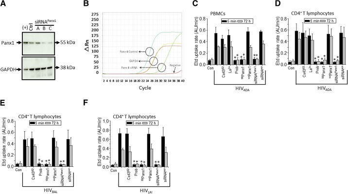
Figure 2. HIV increases the activity of Panx1 hemichannels in human primary CD4+ T lymphocytes.
(A) Total levels of Panx1 in CD4+ T lymphocytes under control conditions (Ctrl) or after transfection for 48 h with three different siRNAs (siRNA A, B, and C) to Panx1 hemichannels were analyzed by Western blot. HeLa cells transfected with Panx1 were used as a positive control of Panx1 protein expression [(+)]. Loading controls, analyzed by probing for GAPDH, are also shown. (B) Representative example of qRT-PCR curves of Panx1 mRNA expression in control (cyan line) and after transfection of siRNA to Panx1 (light green line). GAPDH was used as a control (yellow and red lines). The negative control without enzyme did not show any amplification (dark red line). (C and D) Etd uptake rate for PBMCs exposed to HIVADA (C); CD4+ T lymphocytes exposed to HIVADA (D), HIVBal (E), or HIVLAI (F) after 5 min (black bars) or 72 h (gray bars). (C–F) Control (Con), uninfected conditions are shown. Addition of Cx43 hemichannel blockers, Cx43E2 (1:500 dilution) and La3+ (200 μM), did not affect the Etd uptake rate induced by the virus. In contrast, Panx1 hemichannel blockers, Prob (500 μM) and 10Panx1 (200 μM), completely blocked Etd uptake rate induced by the virus. The negative control using a scrPanx1 peptide (200 μM) did not affect Etd uptake rate induced by the virus. Cells were transfected with siRNA C for Panx1 or with the appropriate siRNAscr (both 10 nM), and Etd uptake rate was analyzed. siRNA for Panx1, but not scramble control, reduced Etd uptake rate induced by the virus. Each value corresponds to the mean ± sd of the Etd intracellular intensity present in at least 20 cells/time-point (n=4; *P<0.005).

