
Figure 2. Pneumonic Clec4d−/− mice exhibit a hyperinflammatory response and severe lung pathology characterized by massive neutrophil accumulation.
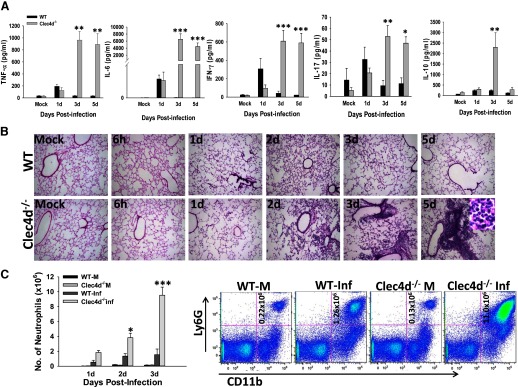
(A) The lungs from mock control and KPn-infected WT and Clec4d−/− mice were harvested at indicated time-points p.i., homogenized in PBS with protease inhibitors, and analyzed for rodent multianalyte profile (Myriad RBM). Results shown are the average of three infected and three mock control mice from three independent experiments. Significant differences are denoted by asterisks (*P<0.05; **P<0.005; ***P<0.001). (B) H&E staining of lung cryosections from mock control and KPn-infected WT and Clec4d−/− mice isolated at indicated times p.i. Original magnification, 100×. The inset in the right panel in the Clec4d−/− row shows a highly magnified area (1000×) of a lesion in infected Clec4d−/−, depicting neutrophils, as indicated by characteristic, multilobed nuclear morphology. (C) Flow cytometry analysis of neutrophils in mock control (WT-M and Clec4d−/− M) and KPn-infected (WT-Inf and Clec4d−/− Inf) mice. Total lung cells were isolated from mice by collagenase treatment at indicated times p.i. The cells were stained with anti-Ly6G-APC and anti-CD11b-Pacific Blue antibodies as markers for neutrophils. Appropriate isotype-matched negative controls were used to set the gates. The bar graph shows the average total number of neutrophils in lungs of three mock control and three KPn-infected WT and Clec4d−/− mice from three independent experiments (total of nine mice/group). Dot plots shown on the right are from one representative mouse in each group. Statistical significance between WT and Clec4d−/− mice is denoted by asterisks (*P<0.05; ***P<0.001).