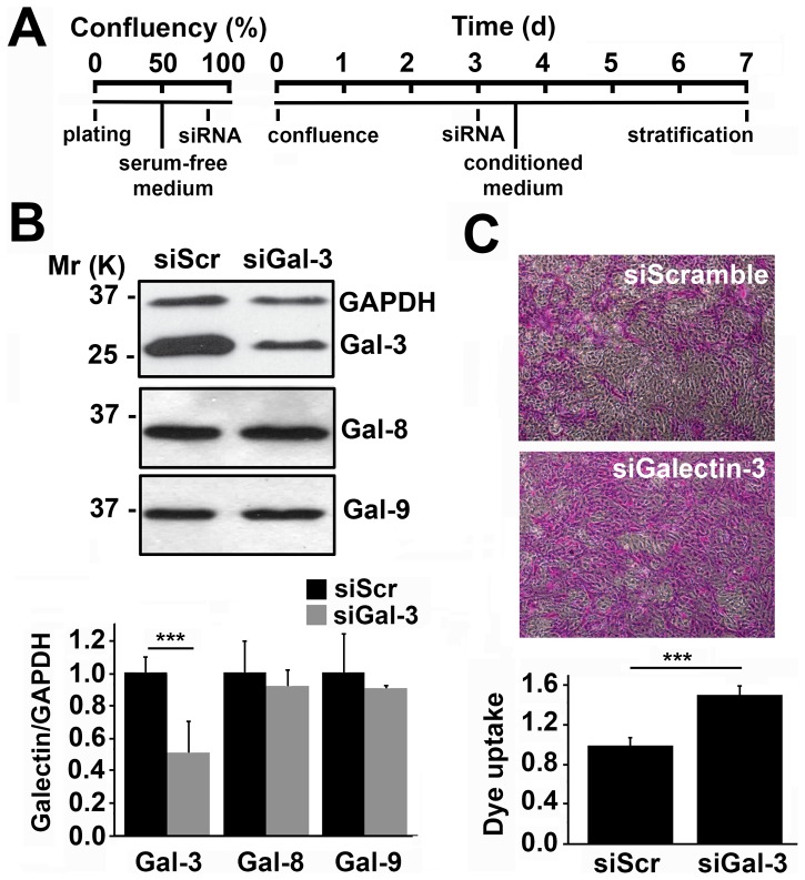
Figure 1. Galectin-3 maintains corneal epithelial barrier function in vitro.
(A) Timeline illustrating the transient abrogation of galectin-3 in a three-dimensional culture system using siRNA. (B) Analyses of whole corneal epithelial cell lysates revealed a 51±18% galectin-3 protein reduction in cultures treated with galectin-3 siRNA (siGal3) as compared to scramble siRNA (siScr). Galectin-3 knockdown did not affect expression of galectin-8 and -9. The upper panel shows representative western blots. (C) The average area of rose bengal staining after galectin-3 knockdown was 51±9% higher than in scramble cells. Representative images for each condition are shown in the upper panel. Images were obtained using a 10× objective lens. All the experiments were performed at least in triplicate and represent the mean ±SD. ***P<0.001.

