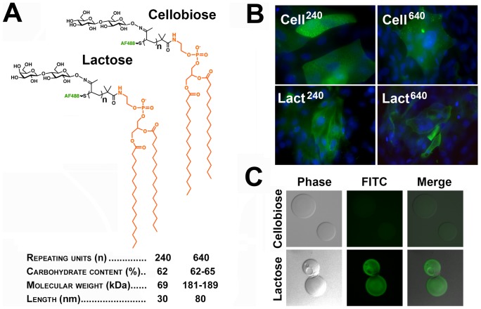
Figure 4. Synthetic glycopolymers incorporate into stratified cultures of human corneal epithelial cells.
(A) Schematic structure and properties of Alexa Fluor 488 cellobiose- and lactose-containing glycopolymers functionalized with a phospholipid end group. (B) Fluorescence microscopy images demonstrated that, following a 1-hour incubation, glycopolymers (green) with 240 and 640 repeating units incorporated into islands of stratified corneal epithelial cells. DAPI was included in the mounting medium to localize the position of the nuclei (blue) in the cell culture. Images were obtained using a 40× objective lens. (C) By pull-down assay, synthetic glycopolymers with lactose-decorated backbones, but not cellobiose derivatives, bound to an rhGal3 affinity column.

