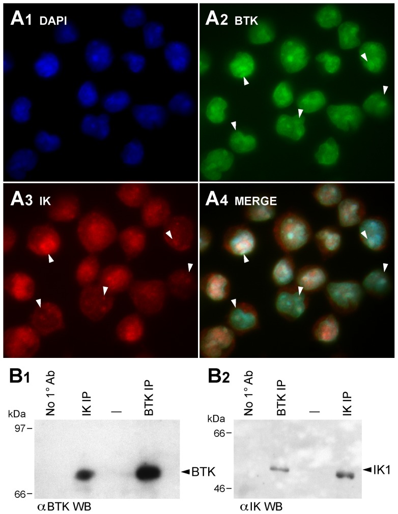
Figure 1. Co-localization and Physical Interactions of Native Ikaros and BTK Proteins in Human Cells.
[A] Nuclear co-localization of Native IK and BTK. ALL-N1 cells were fixed and stained with polyclonal rabbit anti-IK1 (primary Ab)/Alexa Fluor 568 F(ab')2 fragment of goat anti-rabbit IgG (secondary Ab) (red) and mouse anti-BTK MoAb (primary Ab)/Alexa Fluor 488 goat anti-mouse IgG (secondary Ab) (green) antibodies. Nuclei were stained with blue fluorescent dye 4′,6-diamidino-2-phenylindole (DAPI). MERGE panels depict the merge three-color confocal image showing co-localization of IK1 and BTK in DAPI-stained nucleus as magenta immunofluorescent foci (System magnification: 315×). Representative foci of colocalization are indicated with white arrowheads. [B] Co-immunoprecipitation of Native IK and BTK. B1 depicts the results of the BTK Western blot analysis of the IK and BTK immune complexes immunoprecipitated (IP) from ALL-N1 cells. B.2 depicts the results of the IK Western blot analysis of the BTK and IK immune complexes from the same cells. Controls included immunoprecipitations performed without using a primary (10) antibody.

