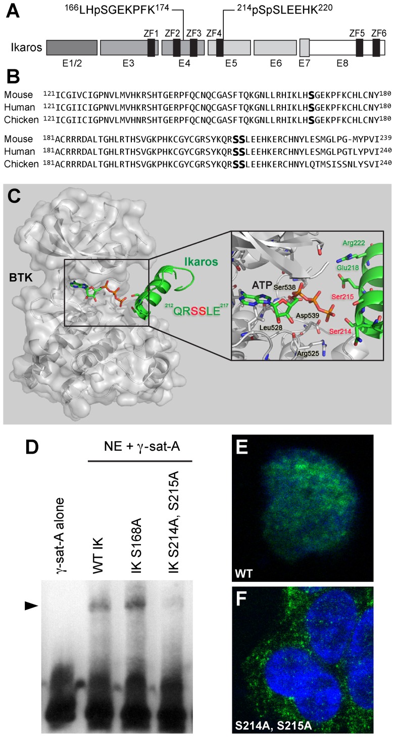
Figure 5. BTK phosphorylation sites of Ikaros.
[A] We identified Ser214 (S214) and Ser215 (S215) as two unique BTK phosphorylation sites within the mouse IK peptide 214SSLEEHK220 corresponding to a consensus sequence encoded by Exon 5 and found in IK from mouse (NM_001025597, human (NM_006060.4), and chicken (NM_205088). [B] Alignment of mouse, human, and chicken IK protein segments containing the identified BTK phosphorylation sites. [C] Schematic diagram of BTK-IK (203–224) complex. BTK is shown as a molecular surface colored in gray, and IK is shown as secondary structure colored in green. Key residues in catalytic site of BTK, ATP and substrate residues (Ser214 and Ser215) in the peptide of IK are shown as a stick model. The model was built by Modeler [1] and shown with Pymol. [D] Loss of IK DNA binding activity by BTK-resistance mutations. EMSAs were performed on nuclear extracts (NE) from Hek293T cells transfected with expression vectors for wildtype (WT) or mutant IK proteins using the Thermo Scientific LightShift Chemiluminescent EMSA Kit and biotin-labeled DNA probe gamma-satellite A. IK activity was measured by the electrophoretic mobility shifts of the biotin-labeled probe, representing IK-containing nuclear complexes (indicated with arrow head). The biotin-labeled DNA was detected using streptavidin-horseradish peroxidase conjugate and a chemiluminescent substrate. The membrane was exposed to X-ray film and developed with a film processor. [E] Confocal two-color merge image of a representative Hek293T cell expressing FLAG-tagged wildtype IK protein (green) in the DAPI-stained (blue) nucleus. (System Magnification: 630×). [F] Confocal two-color merge image of representative Hek293T cells expressing FLAG-tagged mutant IK protein with alanine substitutions at BTK phosphorylation sites S214 and S215 (green) in the cytoplasm outside the DAPI-stained nucleus. (System Magnification: 630×). To detect the FLAG-tagged wildtype (in E) and BTK-phosphorylation site mutant IK proteins (in F), cells were stained with a monoclonal mouse anti-DDK antibody and a secondary goat anti-mouse antibody conjugated with Alexa Fluor 488. Fluorescent cells were imaged using the PerkinElmer Ultraviewer Confocal Dual Spinning Disc Scanner (Shelton, CT).

