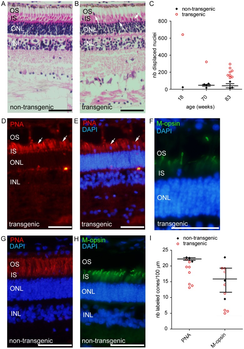
Figure 4. Abnormal retinal morphology in transgenic pigs.
(A) Morphological examination of a retina from a control animal reveals retinal layers: outer segments (OS), inner segment of photoreceptors (IS), photoreceptor nuclei (ONL). (B) Displaced nuclei were observed in the outer segment layer in transgenic retina (arrows). (C) Quantification of the number of displaced nuclei in transgenic and control animals. (D,E,F) Immunolabeling for specific cone markers PNA and M-opsin in transgenic retina identified most of these displaced cells as cones. (G,H) Immunolabeling for specific cone markers PNA and M-opsin in control retina. (I) Quantitation of relative density of displaced cones as determined by PNA or M-opsin labeling across 100 µm on the section. OS: outer segment; IS: inner segment; ONL: outernuclear layer; PNA: peanut agglutinin; M-opsin: medium wavelength opsin; nb: number. Scale bar in A, B and D to F represents 50 µm.

