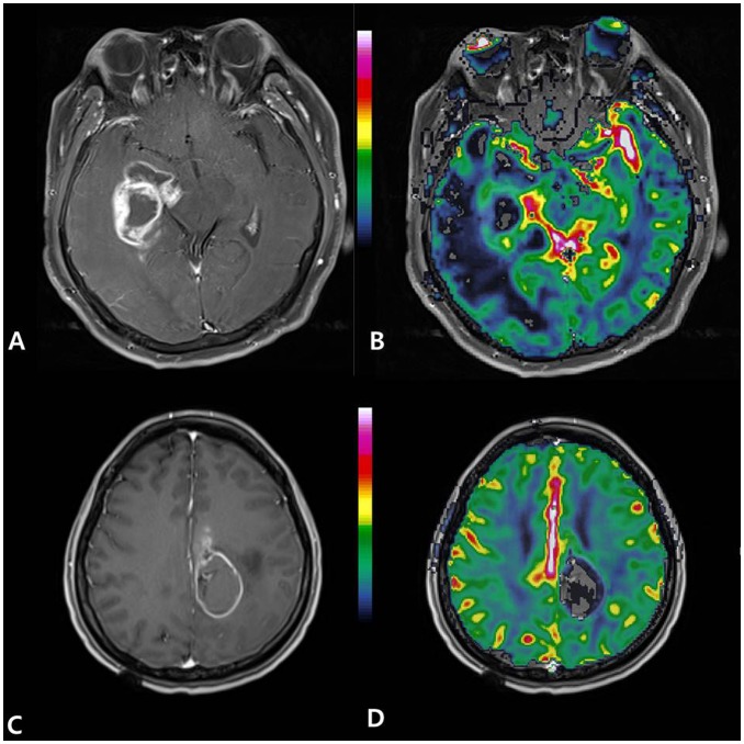
Figure 8. Glioblastomas with favorable genetic profiles show low normalized relative tumor blood volume (nTBV).
(A, B) A 67-year-old man had glioblastoma with EGFR expression (2+), PTEN loss (−), MGMT methylation (+), and a Ki-67 index of 5%. The tumor showed low nTBV (4.3). (C, D) A 36-year-old woman had glioblastoma with EGFR expression (+), PTEN loss (−), MGMT methylation (+), and a Ki-67 index of 9%. The tumor showed low nTBV (4.59) (A, C) Contrast-enhanced T1-weighted axial image, (B, D) normalized relative cerebral blood volume (nCBV) map overlaid on structural contrast-enhanced T1-weighted axial image.

