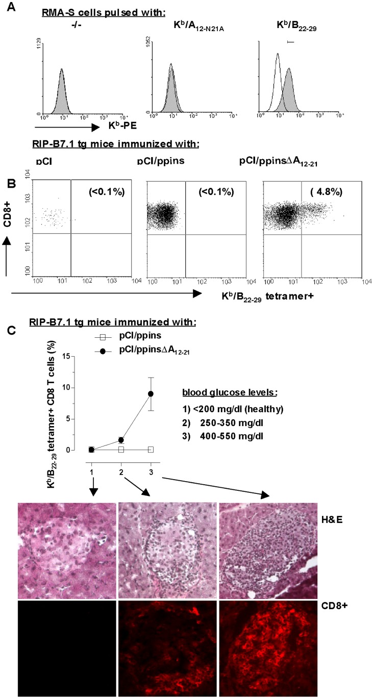
Figure 2. Determination of Kb/B22–29-tetramer+ CD8 T-cells in diabetic RIP-B7.1 tg mice.
(A) TAP-deficient RMA-S cells were either not pulsed (−/−) or pulsed for 6 h with high doses (100 µg/ml) of Kb/A12-N21A or Kb/B22–29 peptides, followed by surface staining of trimeric Kb-molecules and FCM. (B) RIP-B7.1 tg mice were immunized with pCI, pCI/ppins or pCI/ppinsΔA12–21. CD8 T-cells were prepared from pancreata of early diabetic (pCI/ppins, pCI/ppinsΔA12–21) or non-diabetic (pCI) mice and directly stained with Kb/B22–29-tetramers. Primary FACS data are shown for representative mice. The actual percentage of Kb/B22–29-tetramer+ CD8 T-cells within the pancreas-infiltrating CD8 T-cell population is shown in brackets. (C) The numbers of Kb/B22–29-tetramer+ CD8 T-cells were determined during the course of pCI/ppinsΔA12–21-mediated EAD: group 1, health mice (n = 3) with blood glucose levels <200 mg/dl; group 2, early diabetic mice (n = 3) with blood glucose levels between 250–350 mg/dl; group 3, diabetic mice (n = 3) with severe diabetes (i.e., blood glucose levels between 400–550 mg/dl). Pancreata of representative mice out of groups 1 to 3 were analyzed histologically for CD8 T-cell influx (CD8+) or stained with hematoxylin-eosin (H&E).

