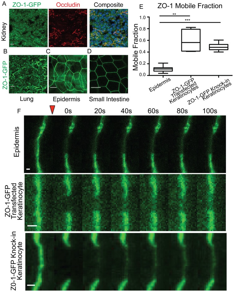
Figure 1. ZO-1 GFP exhibits low mobility in epidermis.
A–D) Localization of ZO-1-GFP in skin sections taken from a ZO-1-GFP knock-in mouse. Scale bar 10 µm. A) ZO-1-GFP co-localizes with the tight junction protein occludin in kidney tissue sections of adult mouse. DNA is stained blue. B) Tissue section of lung taken from adult mouse. C) Whole mount epidermis of embryonic day 17.5 mouse. Note the ZO-1 signal in distinctive cobblestone pattern at cell-cell junctions in the granular layer. D) Whole mount small intestine taken from adult mouse. E) Mobile fractions from FRAP experiments are plotted. The box represents the 25th to 75th percentile and the whiskers represent the 10th and 90th percentiles. ** p<.005. *** p<.0001. F) Representative kymographs are shown of individual FRAP experiments. The bleach point is indicated by the red triangle. Scale bar 1 µm.

