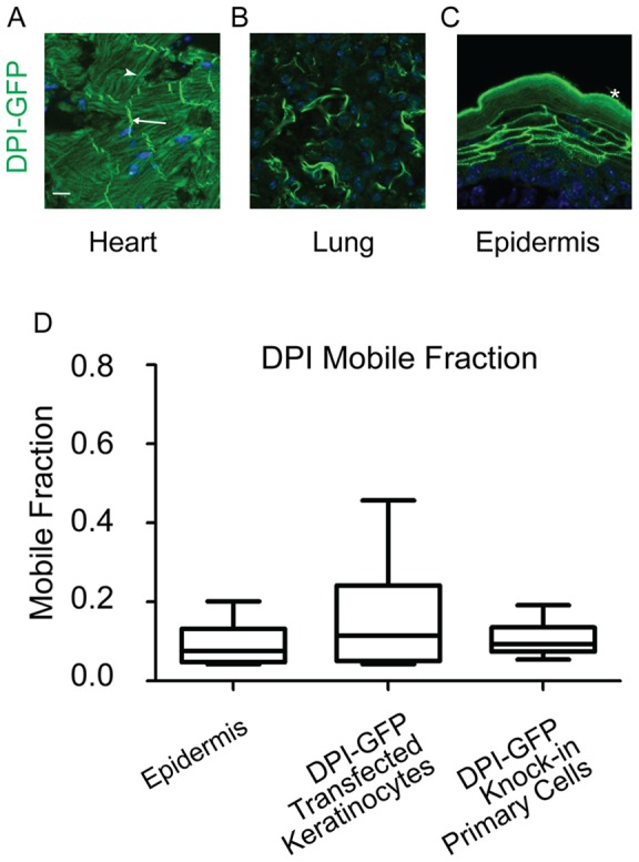
Figure 4. DPI-GFP is stable at desmosomes in both epidermis and in cultured keratinocytes.

A–C) Tissue sections from the adult BAC transgenic DPI-GFP mouse. DNA is stained in blue. Scale bar 10 µm. A) DPI-GFP localization in heart tissue section. Note the strong bands of signal at intercalated discs (arrow). There is strong autofluorescence from thick actin bundles in cardiac muscle fibers (arrow head). B) DPI-GFP localization in lung tissue section. C) DPI-GFP localization in epidermal tissue section. Asterisk labels autofluorescence of cornified layer. D) Mobile fractions from FRAP experiments are plotted. The box represents the 25th to 75th percentile and the whiskers represent the 10th and 90th percentiles.
