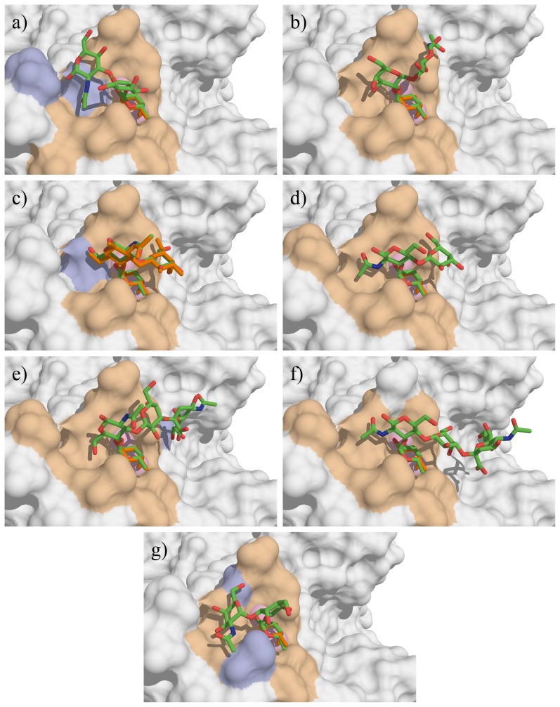
Figure 4. Docking of the oligosaccharides in LecB binding site.
a) H type 1, b) H type 2, c) Lea, d) Lex, e) sLea f) sLex and g) A-tri. The docked oligosaccharides are represented as sticks (carbon, oxygen and nitrogen atoms are colored green, red and blue respectively) and the ones from crystal structures are colored orange. Calcium ions are represented as pink spheres. The protein accessible surface is colored in beige for residues comprised within a sphere of 4 Å around the ligand and in blue for residues involved in hydrogen bond with ligand residues (except fucose).

