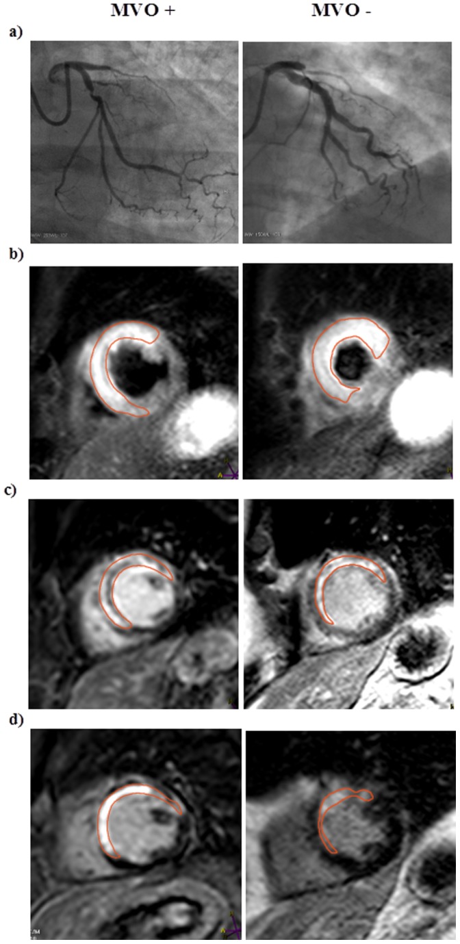
Figure 1. Coronary angiography and CMR images in the presence or absence of microvascular obstruction.

a) Coronary angiography images showing proximal occlusion of the left anterior descending artery, b) CMR demonstrating short axis T2 weighted imaging of myocardium at risk (early CMR), c) short axis late enhancement imaging (early CMR), and d) short axis late enhancement imaging (late CMR), in patients with (MVO +) or without (MVO -) microvascular obstruction. Early CMR was performed 1–4 days after primary PCI and late CMR was performed 4 months later.
