Figure 5.
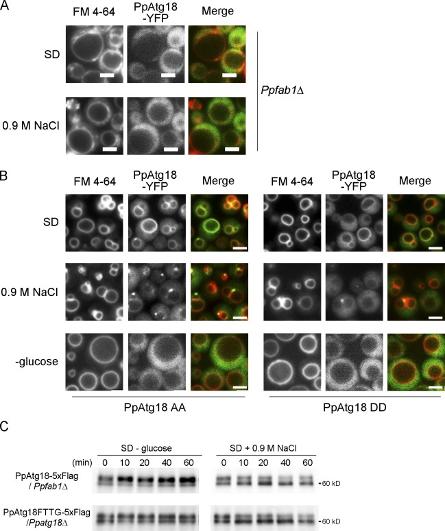
Phosphorylation of PpAtg18 regulates vacuolar membrane dynamics via PI(3,5)P2 binding. (A) Localization of PpAtg18-YFP in Ppfab1Δ. YFP tagged at the C terminus with PpAtg18 was expressed under its own promoter, and vacuoles were stained with FM 4-64. (B) Localization of PpAtg18-YFP phosphorylation mutants. PpAtg18AA, a phosphorylation-defective mutant, and PpAtg18DD, a phosphorylation mimic mutant, appeared similar to the wild type as observed in Fig. 4 B. Bars, 2 µm. (C) Western blot analysis of PpAtg18Wt-5×Flag in Ppfab1Δ or PpAtg18FTTG-5×Flag in Ppatg18Δ.

