Figure 12.

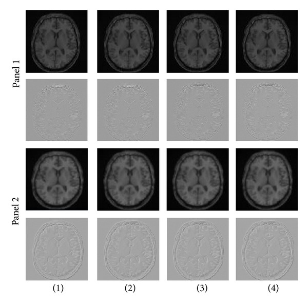
In the top panel one, axial slices from two unregistered 3D MRI volumes using an intensity-based cost function (CR (first column), LS (second column), NCC (third column), and NMI (fourth column)) are shown. Windowed Sinc interpolation was used during registration for the panel one. The second row of the panel one shows subtraction of the images (registered and reference images). The panel two shows similar axial slices from the same two data sets after registration of the full 3D volumes using the same intensity-based cost function (CR (first column), LS (second column), NCC (third column), and NMI (fourth column)). B-spline 3rd-order interpolation was used during registration for the panel two. The second row of the panel two shows subtraction of the images. Although differences of the first rows of panel one and panel two are not easily observed by eye, subtraction of the images shows that differences are present, as seen on the second rows of panel one and panel two.
