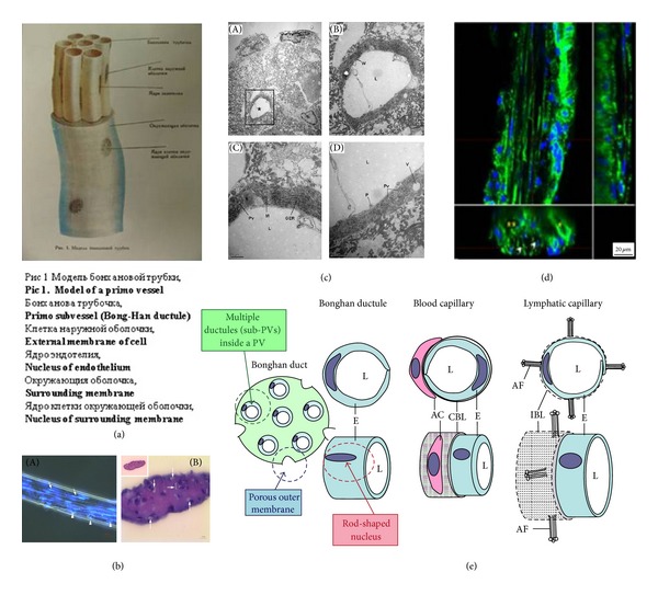
Figure 1.

PV (Bonghan duct) illustrated in various publications. (a) Schematic diagram of a primo vessel, described by Bong-Han Kim with Russian terminologies. English translation of the terminology is added in bold [3]. (b) Optical images showing histological characteristics of a PV. (A) Phase-contrast image of a PV with DAPI. Several sub-PVs (arrow) with rod-shaped nuclei (light blue, arrowhead) are seen. (B) The cross section of a PV: several lumens (arrows) are seen [37]. (c) Electron microscopy of a partial cross section of a PV. (A) A lumen (asterisk) of a sub-PV can be seen. (B) Magnified image of the lumen (rectangular area in (A)). The wall (W) of the sub-PV consists of a single layer of endothelial cells surrounded by fibrin-like fibers. ((C), (D)) Magnified image of (B). L: lumen; M: mitochondria; GER: granular endoplasmic reticulum; P: cytoplasmic protrusion; PV: pinocytotic vesicles; V: vacuole; and F: fibrin-like fiber [37]. (d) Confocal laser scanning microscope image of a PV. The main panel is optical microscopy of the longitudinal section of a PV (the middle section, cells with rod-shaped nuclei in blue), accompanied by a venule and an arteriole on each side (cells with circular nuclei). The lower panel is a cross section of the PV, showing multiple lumens (open arrowheads), and the venule (asterisk) and the arteriole (two asterisks) on its each side. The PV diameter was approximately 30 μm [33]. (e) Anatomical comparisons of characteristics of the PV, blood, and lymphatic capillaries. The PV has multiple sub-PVs within, and the PV's outermost layer has large pores. The endothelial cells of the sub-PV possess rod-shaped nuclei. E: endothelium; L: lumen; AC: accessory cell; CBL: complete basal lamina; IBL: incomplete basal lamina; and AF: anchoring filaments [37].
