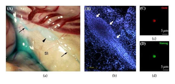
Figure 2.

Images of PVS. (a) An image of a PN (Bonghan corpuscle; arrowhead) found on the rabbit small intestine, with PVs (arrows) at both ends, using methylene blue as the contrast agent [37]. (b) An image of a PN (arrows), which was identified lower part of the superior sagittal sinus of a rabbit brain [33]. DAPI staining of the nuclei of the cells inside the PN. Very small cells are packed in the PN. ((c), (d)) Immunostaining of the small cells isolated from PNs on the surfaces of rat intestine, for the embryonic stem cell markers Oct4 (red) and Nanog (green). The scale bar indicates 5 μm [38].
