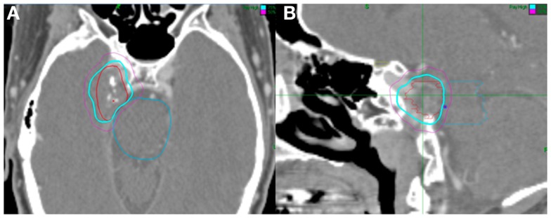
Figure 1.

Fifty-three-year-old man with a right Meckel cave meningioma. He presented with right facial pain. The decision was to proceed with radiosurgery. Treatment planning axial (A) and sagittal (B) computed tomography images demonstrating the GTV (red), brainstem (blue), and chiasm (yellow). Isodose lines shown as follows: blue 79% (prescription) and purple 50%. Note proximity of the meningioma to the brainstem. The tumor was treated with 2500 cGy in five fractions and his pain resolved.
