
Fig. 2.
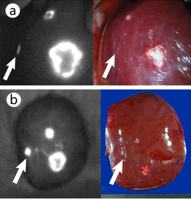
ICG fluorescence imaging on liver surfaces (left) and their gross appearances (right) (from [21]). a Fluorescence imaging prior to liver resection enabled visualization of the metastasis of colorectal cancer that was palpable but grossly unidentifiable (arrow), as well as the other two lesions visible on the liver surface. b Fluorescence imaging of the resected specimen. The arrow indicates a grossly unidentifiable tumor that was located 0.8 cm below the liver surface.