
Fig. 12.
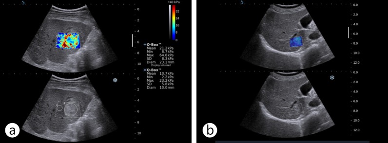
US elastography images of HCC and hemangioma. A hyperechoic mass was shown to have increased stiffness on US elastography using SSI (a). It was confirmed as HCC on biopsy. A small hyperechoic low-stiffness nodule with a stiffness value similar to that of liver parenchyma on US elastography using SSI (b) was revealed as a hemangioma on follow-up MR.