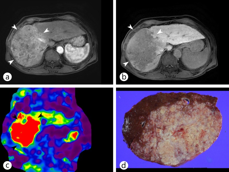
Fig. 13.
MR elastography of a surgically confirmed huge infiltrative HCC. a, b On Gd-EOB-DTPA-enhanced MR images during the arterial (a) and portal (b) phases, the main mass (arrowheads) showed the typical enhancing pattern of HCC, with arterial enhancement and washout, in right lobe of the liver. c MR elastography showed the increased stiffness of the tumor compared with the adjacent liver parenchyma. d The gross specimen showed a large mass with an infiltrative growth pattern.

