Fig. 8.
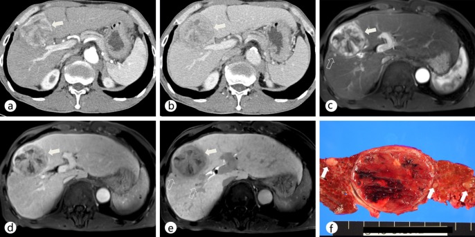
Comparison of dynamic MDCT and Gd-EOB-DTPA-enhanced MRI in a patient with a dominant hypervascular HCC in segment VIII as well as daughter nodules. a, b On contrast-enhanced CT scans during the arterial (a) and portal (b) phases, the main mass (arrow) showed arterial hyperenhancement and washout on the portal and delayed phases in the right lobe of the liver. c, d On Gd-EOB-DTPA-enhanced MR images during the arterial (c) and portal (d) phases, the main mass (arrow) shows a typical enhancing pattern of HCC in the right lobe of the liver. e On a hepatobiliary-phase image, two additional daughter nodules (open arrows) that were not detected on a CT scan were seen as hypointense nodules. f The gross specimen shows several additional daughter nodules (open arrows) around the main tumor.

