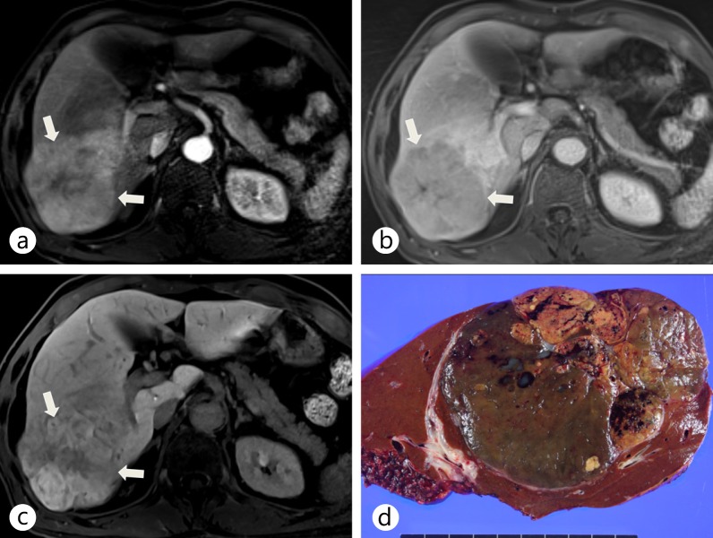
Fig. 9.
Surgically confirmed HCC showing increased contrast uptake on hepatobiliary-phase imaging of Gd-EOB-DTPA-enhanced liver MRI. a An arterial-phase Gd-EOB-DTPA-enhanced liver MRI shows a hyperenhancing tumor (arrows) in the right posterior segment of the liver. b A portal-phase Gd-EOB-DTPA-enhanced liver MRI shows a hypointense lesion (arrows). c On a hepatobiliary-phase image, the tumor (arrows) shows hyperenhancement resulting from increased expression of OATP8, probably due to genetic alterations. d The gross specimen shows a large mass with mixed components and bile staining.

