
Figure 2. Leptin receptor (OBR) protein expression is restricted to thymic medullary epithelial cells.
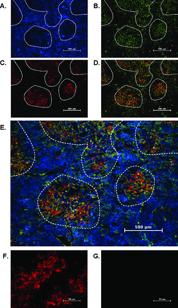
Frozen thymus sections from 12 week old mice were stained with an mTEC marker, cytokeratin 5 (K5) (green), and leptin receptor (pan marker, red). Nuclei were stained with DAPI (blue). Panels depict staining for DAPI (A), cytokeratin 5 (B), leptin receptor (C), leptin receptor and K5 overlay (D) and leptin receptor and K5 overlay with DAPI (E). Final magnification 40X. Areas of medulla are outlined by dashed line. Final magnification 400X shows leptin receptor (red) (F) or isotype control antibody (G). Representative section from five mice per group.