
Figure 5. Leptin-mediated protection of thymic stroma during LPS challenge.
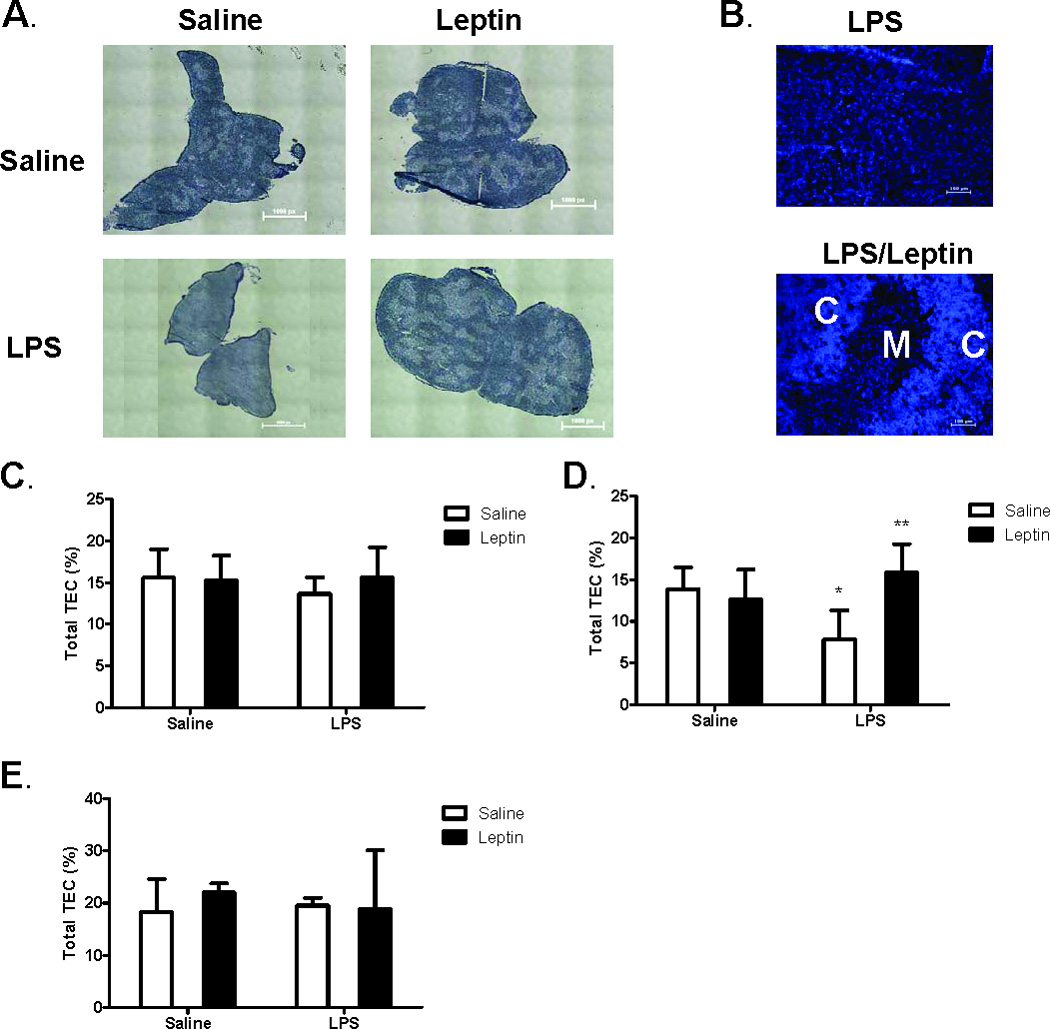
Female C57BL/6 mice were administered either saline or leptin (1 µg/g body weight, IP) and simultaneously challenged with either saline or E. coli LPS (100 µg/mouse, IP). Hematoxylin and Eosin staining of representative thymus sections (A) seven days post treatment. Whole tissue stitched image, total magnification (40X). Calibration 1.61 µm equals 1.0 pixels. DAPI staining of thymocyte nuclei (B) depict distinct areas of cortex (C) and medulla (M). Total magnification (100X). Frequency of thymic epithelial cells (MHC Class II+ cells) in CD45- stromal cell population one day (C), three days (D), and seven days (E) post treatment. Data presented are mean ± SD from four mice per group. * p ≤ 0.05 compared with saline-treated controls. ** p ≤ 0.05 compared with LPS-treated controls.