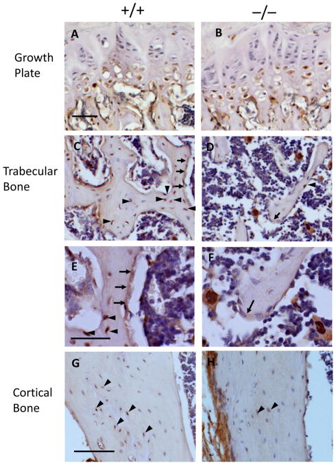
Fig 1. Expression of IGF-1R in Igf-1r−/− mice.
Immunohistochemistry staining showed that in the growth plate (A & B), IGF-1R (brown) was expressed in the prehypertrophic and hypertrophic chondrocytes in the Igf-1r+/+ (A) mice and the Igf-1r−/− (B) mice; in the trabecular bone of the secondary spongiosa, IGF-1R is expressed in the osteoblasts lining the bone surfaces (arrows) and osteocytes embedded in the bone matrix (arrowheads) in the Igf-1r+/+ mice (C), but very little expression of IGF-1R was identified in the osteoblasts (arrows) and osteocytes (arrow heads) in the Igf-1r−/− mice (D). High magnification pictures of Igf-1r+/+ (E) and Igf-1r−/− (F) mice are shown below. In the cortical bones, IGF-1R is expressed in the osteocytes (brown, arrow heads) in the Igf-1r+/+ mice (G), but very few osteocytes (arrowheads) express IGF-1R in the Igf-1r−/− mice (H). 20 X in A–D, G & H; 40 X in E & F. Bars = 50 μm.

