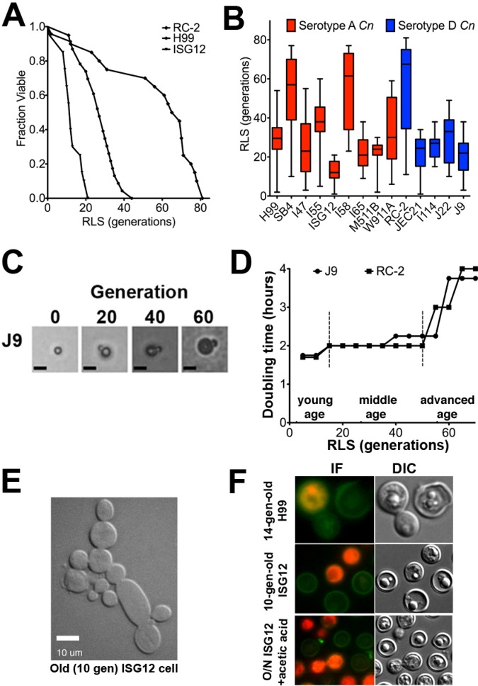
FIG 1 .
Clinical C. neoformans strains demonstrated variability in their life span. (A) Recording of RLS of individual cells of C. neoformans by generation of survival curves demonstrated short (ISG12), medium (H99), and long (RC-2) RLSs of strains that were statistically different from each other (P < 0.01 by Wilcoxon rank sum test). (B) The median RLSs (short horizontal lines in the boxes) of clinical serotype A and D C. neoformans strains were highly variable (20 to 60 cells per strain). The minimum and maximum values for all data are indicated by the ends of the whiskers of the box plots. (C) Increasing cell size was observed during the RLS shown for C. neoformans strain J9 at 0, 20, 40, and 60 replications. Bars, 10 µm. (D) The RLS of C. neoformans strains (J9 and RC-2) can be divided into young, middle, and advanced age based on changes in DT. (E) 10-generation-old C. neoformans cells (ISG12) manifested abnormal budding. Bar, 10 µm. (F) In order to investigate how cells of advanced age died, 14-generation-old H99 cells or 10-generation-old ISG12 cells were stained with annexin V. Cells undergoing apoptosis stained positive for annexin V (green), those undergoing necrosis stained for propidium iodine (PI) (red), and the cells that had suffered loss of membrane integrity stained for both (red with green boundary). Young cells (overnight [O/N] culture) were treated with acetic acid to induce apoptosis and stained similarly. The cells were examined by immunofluorescence (IF) and differential interference contrast (DIC) microscopy.

