
Figure 1.
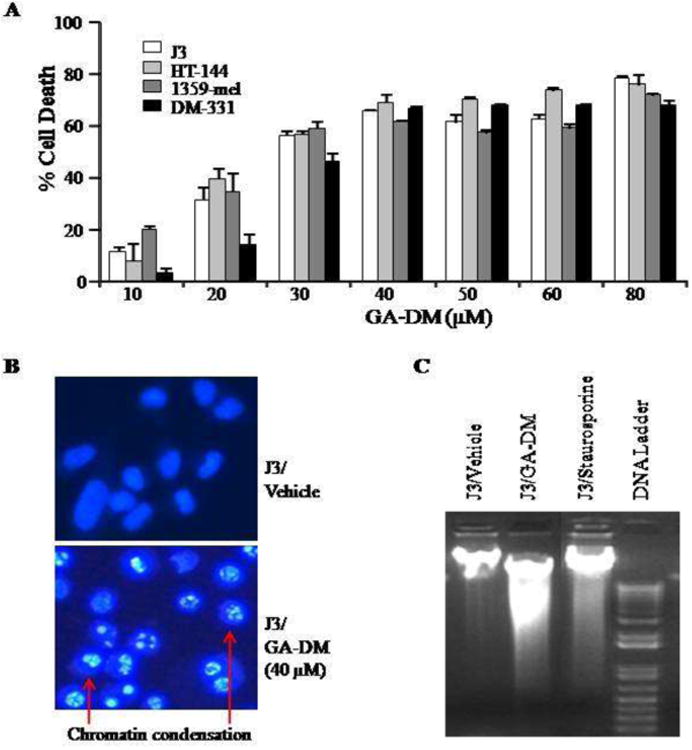
Anti-proliferative and apoptotic activity of GA-DM on human melanoma cells. (A) Cells were treated with vehicle (DMSO) alone or GA-DM (10-80μM) for 24h at 37°C, followed by the MTS viability assay as described in the Materials and Methods. Control cells treated with vehicle alone were utilized to calculate the percent cell death induced by GA-DM as indicated. The data shown are results of at least three separate experiments that were performed in triplicate wells. Error bars represent mean ± S.D. (B) Hoechst staining of J3 melanoma cells treated with vehicle alone or 40 μM of GA-DM. Cells treated with GA-DM showed typical morphology of the apoptotic nuclei stained with Hoechst, in which chromatin was condensed and aggregated at the nuclear membrane as indicated by bright fluorescence at the periphery (arrows). (C) Agarose gel electrophoresis of DNA extract showing internucleosomal DNA fragmentation after treatment of J3 cells with GA-DM (40μM) for 24 h. Cells treated with staurosporine (1 μM) was used as positive control.