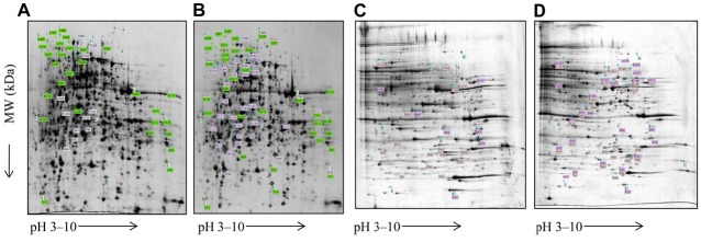
Figure 1. Cellular and membrane protein spots of C glabrata strains that were resolved using two-dimensional gel electrophoresis. Spots representing differentially expressed proteins were later identified by liquid chromatography-tandem mass spectrometry (LC-MS/MS) peptide mass fingerprinting. (A) Cellular protein spots of fluconazole-susceptible strains. (B) Cellular proteins spots of fluconazole-resistant strains. (C) Membrane protein spots of fluconazole-susceptible strains. (D) Membrane protein spots of fluconazole-resistant strains.

