Figure 1. .
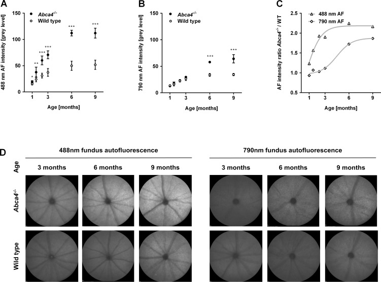
(A–C) Longitudinal measurements of fundus AF intensity in the Abca4−/− mouse (n = 5) and WT (n = 8) controls between 1 to 9 months of age. To distinguish lipofuscin- and melanin-related fundus AF, 488 (A) and 790 nm (B) wavelengths were used for excitation, respectively. Levels of fundus AF (mean ± SD) were significantly different between strains and between age groups (P < 0.01, two-way ANOVA) for 488 and 790 nm fundus AF. *P < 0.05; **P < 0.01; ***P < 0.001. (C) Using the ratio of mean fundus AF between Abca4−/− and WT mice controlled for age-related factors potentially affecting AF intensity measures. Curves were fitted to illustrate the time course of AF increase in Abca4−/− relative to WT at the two different excitation wavelengths. (D) Unprocessed representative recordings of fundus AF excited at 488 and 790 nm in Abca4−/− and WT control mice (cross-sectional cohort). Overall, the AF signal was higher with 488 nm compared with 790 nm excitation light. At 3, 6, and 9 months, 488 nm AF in Abca4−/− mice is higher than in WT, with a larger difference in older animals. 790 nm AF was only significantly different in 6 and 9 months old animals, where Abca4−/− mice show a higher AF level than controls.

