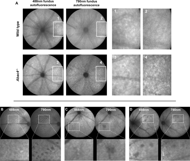
Figure 2. .
(A) Representative AF fleck patterns in a 12-month-old Abca4−/− mouse and an age-matched control (images processed for contrast). Flecks on 488 nm AF images were visible in both mice but were more pronounced in the Abca4−/− mouse. On 790 nm AF, a fleck pattern was only visible in the Abca4−/− mouse, but not in the WT control. (B–D) Dark areas on fundus AF imaging suggesting focal damage of the RPE in aged Abca4−/−mice. (B, C) 488 nm fundus AF (left) in a 12- (B) and 18- (C) month-old mouse showing fleck-like increased AF and faint spots of reduced AF. The latter are also hypofluorescent on the 790 nm AF image (right). (D) Rarely, spots of markedly reduced AF were more obvious on 488 nm AF images. These lesions were not seen in similarly aged WT mice.

