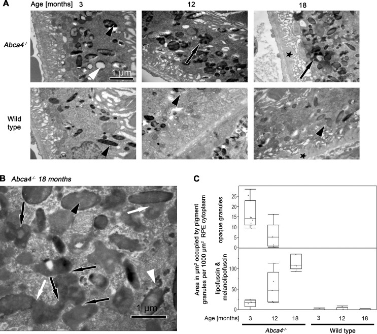
Figure 6. .
(A) TEM micrographs of RPE cells of from Abca4−/− and WT mice at ages 3, 12, and 18 months. In 3-month-old Abca4−/− mice electron-opaque homogeneous granules are labeled by a white and a melanosome by a black arrowhead. The granule labeled by the white arrowhead represents the more classical type of lipofuscin. With progression of age, unusual granules of irregular shape and electron density (black arrows) accumulate in the RPE cytoplasm of 12- and 18-month-old Abca4−/− mice. This type of organelle is nearly absent from WT mice. Melanosomes are indicated by black arrowheads. Eighteen-month-old mice of both groups contain electron opaque material (asterisks) between Bruch's membrane and basal infoldings. (B) TEM micrograph of the RPE from an 18-month-old Abca4−/− mouse at high magnification. With progression of age, unusual granules accumulated in the cytoplasm of RPE cells. A melanosome is marked by an arrowhead and Bruch's membrane by an asterisk. The material accumulated in the cytoplasm is irregular in shape and electron dense (black arrows). These granules appear to fuse with each other (black arrows) and with melanosomes (white arrows). The electron density of these confluent granules is occasionally as dense as in melanosomes (black arrowhead). A melanosome in a state of disintegration is indicated by a white arrowhead and were also present in WT mice. (C) Quantification of lipofuscin and melanolipofuscin granules by electron microscopy. The total areas (μm2) occupied by lipofuscin and melanolipofuscin per 1000 μm2 sectioned RPE cytoplasm increased significantly in Abca4−/− mice at 12 and 18 months compared with age-matched WTs. The area occupied by the classic (opaque) lipofuscin granules in Abca4−/− mice declined with age.

