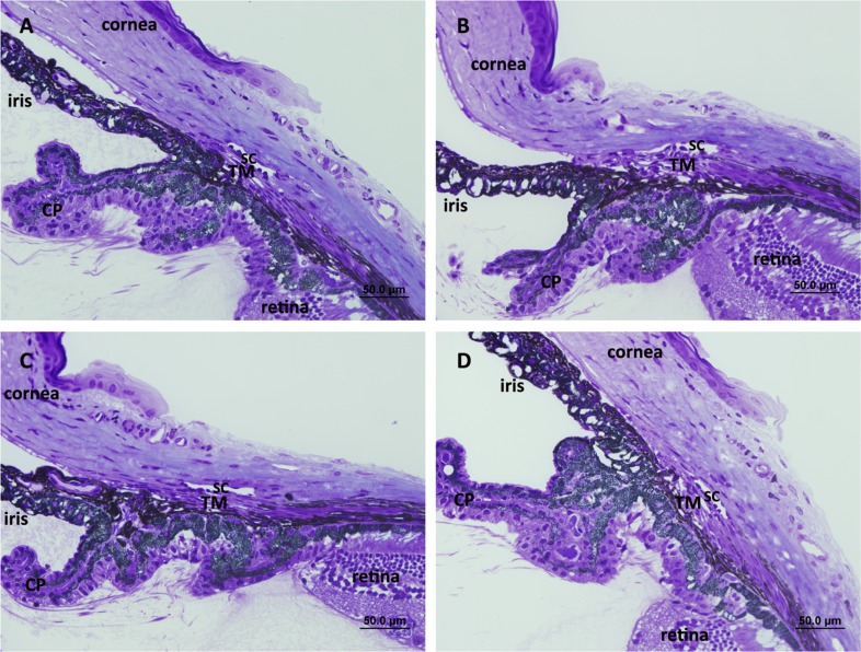
Figure 7. .
Light microscopic images of iridocorneal angles of (A) TNC wild-type, (B) TNC-null, (C) TNX wild-type, and (D) TNX-null mice. Schlemm's canal (SC), trabecular meshwork (TM) beams and cellularity, uveoscleral outflow pathway, and ciliary body location appeared grossly indistinguishable between TNC wild-type and TNC-null and between TNX wild-type and TNX-null mice, respectively. CP, ciliary processes. Scale bar: 50 μm.

