
Fig. 8.
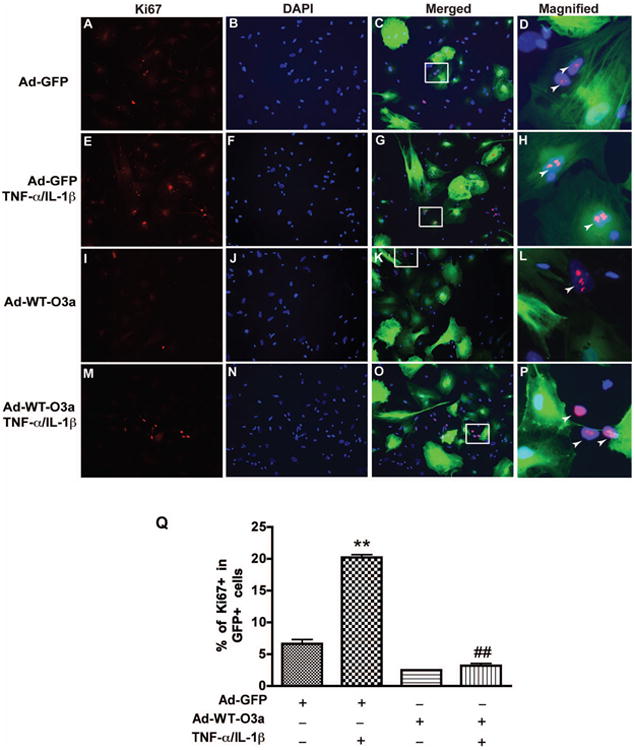
Overexpression of wild type of FOXO3a inhibits cytokine-mediated proliferation of human astrocyte. After 24 h of adenovirus infection, cells were treated with TNF-α and IL-1β for 3 days and then stained with Ki67. DAPI (blue) was used as a nuclear marker to count the total cell number. A-C, Ad-GFP infection without TNF-α and IL-1β treatment. E-G, Ad-GFP infection with TNF-α and IL-1β treatment. I-K, Ad-WT-FOXO3a infection without TNF-α and IL-1β treatment. M-O, Ad-WT-FOXO3a infection with TNF-α and IL-1β treatment. C, G, K, and O are KI67, DAPI and GFP merged images of each group. D, H, L, and O are magnified Images from outlined square from C, G, K, and O respectively. Arrow indicates the Ki67+/GFP+ cells in D and H, Ki67+/GFP- cells in L and O. Q, quantification of three donors. **, p < 0.01, compared with Ad-GFP without TNF-α and IL-1β treatment; ##, p < 0.01, compared with Ad-GFP in TNF-α and IL-1β treatment.