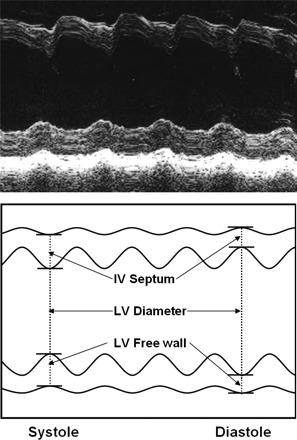
Fig. 1.

An actual M-mode recording (top) with a schematic diagram (bottom). The measurement of septal and anterior wall thickness and chamber diameter in systole and diastole are shown. Similar data were obtained during standing and sitting, during leg lifting, and during exercise. IV, intraventricular; LV, left ventricular.
