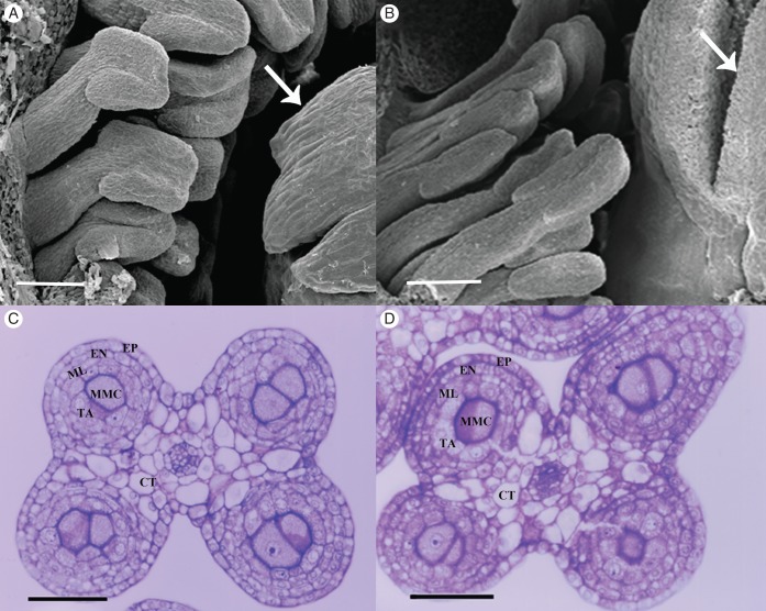
Fig. 2.
Stamens of male and female flowers at stage 5. (A) Detail of a male flower viewed by scanning microscopy showing stamens and pistil (arrow). (B) Detail of a female flower viewed by scanning microscopy showing stamens and pistil with a differentiated stigmatic lobe (arrow). (C) Cross-section of a young tetrasporangiate anther of a male flower bud at stage 5. (D) Cross-section of a young tetrasporangiate anther of a male flower bud at stage 5. Abbreviations: EP, epidermis; EN, endothecium; ML, middle layer; TA, tapetum; MMC, microspore mother cells; CT, connective tissue. Scale bars: (A, B) = 200 µm; (C, D) = 50 µm.

