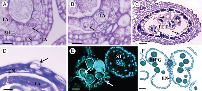
Fig. 5.

Male flower anther wall development. (A, B) Different stages of karyokinesis. (B–F) Consecutive stages in anther wall development. (A) The young anther wall is composed of the epidermis (EP), endothecium (EN), middle layer (ML) and tapetum (TA). The tapetal cells undergo karyokinesis at stage 6; the arrow shows the chromosomes migrating at anaphase. (B) Tapetal cells are binucleate (arrow) by the time the MMCs undergo meiosis during stages 6 and 7. (C) At stage 8, the tetrad are surrounded by callose, the tapetal cells become densely cytoplasmic and Ubisch bodies (structures of sporopollenin involved in pollen wall formation) are released into the locule (arrow); debris of the ephemeral middle layer remain. (D) The tapetum continues degenerating and has a granular appearance, while the endothecium and epidermis show shrunken cytoplasm; some cells of the epidermis have a conical appearance (arrow). (E) At stage 10, anthers stained with DAPI show DNA content in epidermal cells (right arrow), pollen grains (left arrow) and in the filament of the stamen. Autofluorescence was detected in the fibrous thickenings in the endothecium and in the pollen grain wall. (F) Anther, containing mature pollen grains, showing ruptured stomium (arrow) and fibrous thickening in the endothecium. Abbreviations: EN, endothecium; ML, middle layer; TA, tapetum; MMC, microspore mother cell; TET, tetrad; MPG, mature pollen grain; ST, stamen. Scale bars: (A, B) = 10 µm; (C) = 30 µm; (D) = 5 µm; (E) = 60 µm; (F) = 100 µm.
