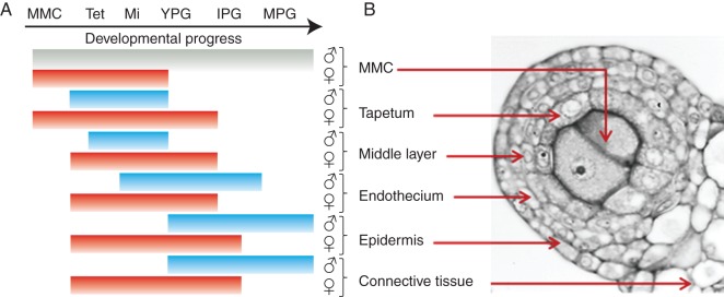
Fig. 9.

Spatial and temporal hallmarks of PCD in anther tissues of male flowers (blue lines and ♂) and female flowers (red lines and ♀). The lack of PCD in the MMCs in male flowers is represented by a grey line. (A) Developmental stages are represented as: MMC, microspore mother cell in pre-meiotic stage; Tet, MMC undergoing meiosis to microspore tetrad; Mi, callose-released microspore; YPG, young pollen grain; IPG, intermediate pollen grain; MPG, mature pollen grain. (B) Cross-sections of an anther at the MMC stage showing the sporophytic and gametophytic tissue.
