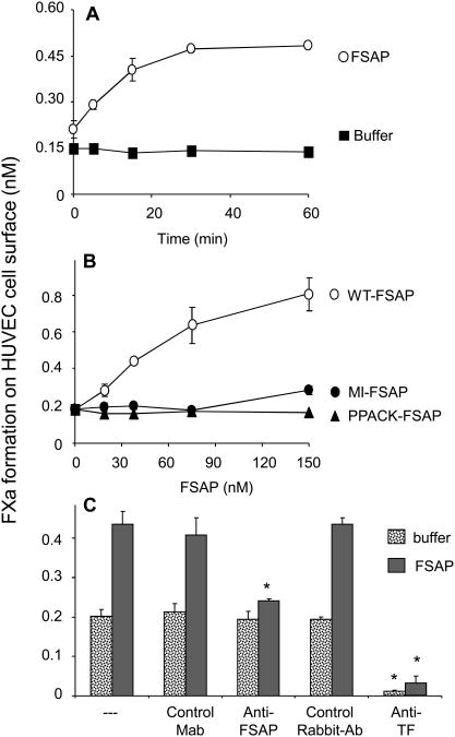
Fig. 1. Effect of FSAP on FXa generation on HUVEC surface.
(A) Cells were activated for 6h with TNFα (25ng/ml) and then treated with FSAP (150 nM) (O) or its control buffer (■) for the indicated times and FXa generation was measured. (B) In TNFα-activated cells the effect of increasing concentrations of WT-FSAP (O), MI-FSAP (●) and PPACK-FSAP (▲), added for 60 min, on FXa generation was measured. (C) In TNFα-activated cells FSAP (150 nM) was added for 60 min and FXa was measured in the presence of an anti-TF Ab or a control Ab or anti-FSAP Mab (#570) or control Mab (all 20 μg/ml). In panels A-C results are shown as mean ± SD (n=3). * indicates a statistical significance p < 0.05.

