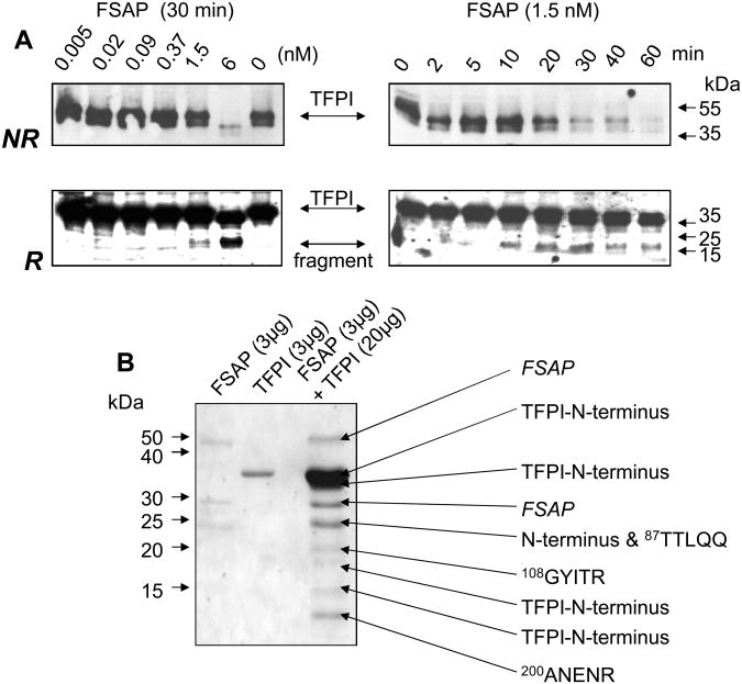
Fig. 5. Characterization of the cleavage of recombinant TFPI by FSAP.
(A) Cleavage of recombinant TFPI by FSAP was followed by Western blotting. In the left panels TFPI (14 nM) was incubated with increasing concentrations of FSAP (0-6 nM) as indicated for 30 min. Western blots were performed with an anti-TFPI polyclonal antibody under non-reducing conditions (NR) or after reduction (R) of samples. In the right panels TFPI (14 nM) was incubated with FSAP (1.5 nM) for the indicated times and Western blots were performed. Migration of MW markers and TFPI as well as a degradation fragment are indicated with arrows. (B) Determination of the cleavage sites in TFPI. TFPI and FSAP were incubated for 30 min at 37°C as indicated and run on SDS-PAGE and blotted on to a PVDF membrane. This was stained with Coomassie blue and the protein bands were subjected to amino terminal sequencing using an automated Edman sequencer (Applied Biosystems). Numbers refer to position of amino acid in full length mature TFPI protein.

