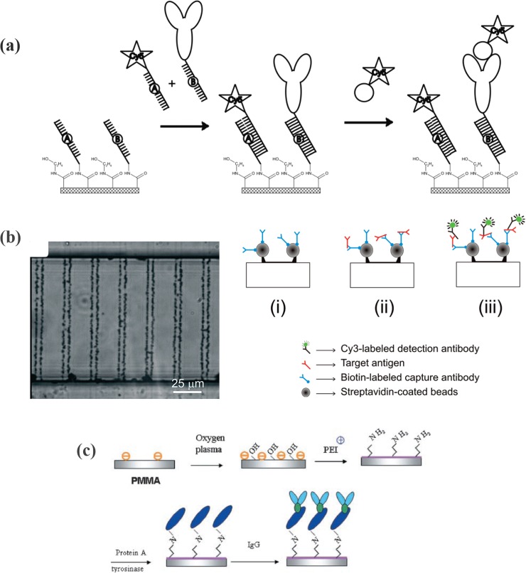
Figure 14.
(a) Sequence-specificity control after immobilization of two different receptor sequences (A and B) onto the COC surface. First, Cy5-labeled oligonucleotide complementary to sequence A and afterward rabbit-IgG conjugated oligonucleotide complementary to sequence B were hybridized. Reprinted with permission from H. Wang et al., Electrochem. Commun. 12, 258 (2010). Copyright 2010 Elsevier. (b) Optical micrographs of double-line bead micropatterns and schematic illustration of the sandwich immunocomplex formation on the streptavidin-coated bead micropattern. The microchannel is filled and incubated, subsequently, with (i) biotin-labeled capture antibody, (ii) target antigen, and (iii) Cy3-labeled detection antibody, forming the sandwich immunocomplex. Reprinted with permission from V. Sivagnanam et al., Anal. Chem. 81, 6509 (2009). Copyright 2009 American Chemical Society. (c) Schematic illustration of the stepwise process involved in the TR-catalyzed protein A-based antibody immobilization. Reprinted with permission from Y. Yuan et al., Biotechnol. Bioeng. 102, 891 (2009). Copyright 2008 Wiley InterScience.

