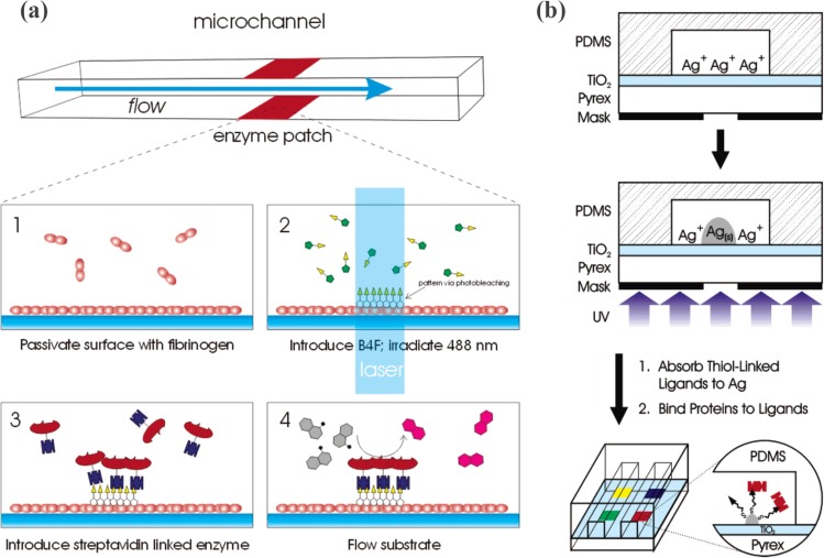
Figure 16.
(a) Schematic diagram of the photoimmobilization process of enzyme. (1) Passivation of the surface with a fibrinogen monolayer is followed by (2) biotin-4-fluorescein surface attachment. This is accomplished by photobleaching with a 488-nm laser. (3) Next, the binding of streptavidin-linked enzymes that can be exploited to immobilize catalysts and (4) monitor reaction processes on-chip. Reprinted with permission from M. A. Holden et al., Anal. Chem. 76, 1838 (2004). Copyright 2004 American Chemical Society. (b) Schematic diagram for the protein patterning using a silver nanoparticle film. First, an AgNO3 solution is introduced into the microchannel. Next, UV radiation is passed through a photomask onto the backside of the TiO2 thin film. Ag+ ions adsorbed at the interface are selectively reduced by photoelectrons, which grow into nanoparticle films. Thiol chemistry was used to immobilize streptavidin. Reprinted with permission from E. T. Castellana et al., Anal. Chem. 78, 107 (2006). Copyright 2006 American Chemical Society.

