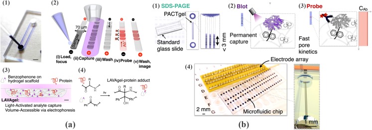
Figure 18.
(a) Design and operation of the microfluidic IEF assay. (1) Glass microfluidic device (scale bar: 2 mm), (2) the 80-min five-step assay is completed in a single microchannel, (3) schematic of microchannel cross-section depicting photoactivated protein immobilization: analytes are electrophoresed through the PA gel, exposed to UV, and covalently immobilized (scale bar: 5 μm), (4) schematic of reaction between polypeptide backbone and benzophenone copolymerized in the PA gel. Ph denotes phenyl group. Reprinted from permission from A. J. Hughes and A. E. Herr, Proc. Natl. Acad. Sci. U.S.A. 109, 5972 (2012). Copyright 2012 National Academy of Sciences, USA. (b) Single-channel microfluidic Western blotting. The microfluidic Western blotting step is comprised of: (1) analyte stacking and SDS-PAGE within the PA gel; (2) capture of separated protein bands (“blotting”) onto the benzophenone-copolymerized PA gel under UV exposure; (3) electrophoretic introduction of fluorescently labeled detection antibodies for the target analyte; and (4) standard microscope-slide-sized chips with a scalable electrode array, accommodating 48 blots per chip in triplicate (144 microchannels). Reprinted from permission from A. J. Hughes and A. E. Herr, Proc. Natl. Acad. Sci. U.S.A. 109, 21450 (2012). Copyright 2012 National Academy of Sciences, USA.

