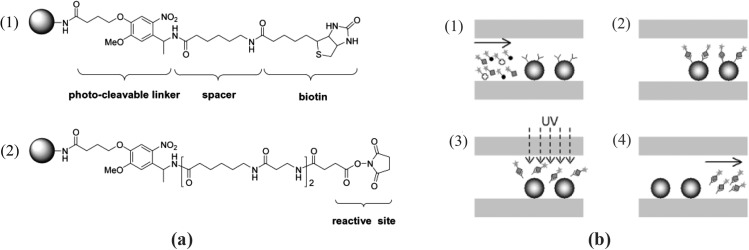
Figure 20.
(a) Structures of the photo-cleavable sites on the bead. (1) Biotinylated bead with a short spacer and (2) active ester containing bead with a long spacer for aptamer coupling. (b) Schematic view of microaffinity purification process. (1) Injection of the protein mixture into the microchip packed with microbeads, (2) purification of the target protein, (3) UV irradiation, and (4) analysis of the photolytically eluted protein. Reprinted with permission from W. J. Chung et al., Electrophoresis 26, 694 (2005). Copyright 2005 Wiley InterScience.

