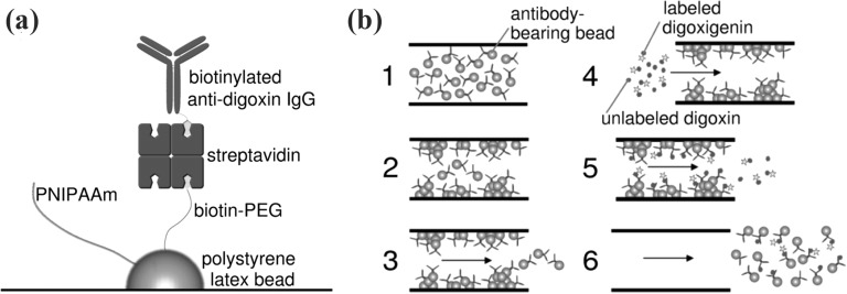
Figure 21.
Schematic representations of the temperature-responsive bead immunoassay system. (a) A 100 nm diameter latex nanobead is surface-conjugated with biotin–PEG and PNIPAM. Streptavidin is bound to the exposed biotin, providing binding sites for the biotinylated anti-digoxin IgG, and (b) a schematic of the experimental protocol. (1) Suspended beads are loaded into the microfluidic channel, (2) the temperature in the channel is then increased from room temperature to 37 °C, resulting in aggregation and adhesion of the beads to the channel wall, (3) flow is initiated, washing unadsorbed beads out of the channel, (4) a mixture of fluorescently labeled digoxigenin and digoxin is flowed into the channel, (5) components of this mixture that fail to bind the immobilized antibodies are washed through, and (6) finally, the temperature in the channel is reduced, and the aggregation-absorption process is reversed as antigen-bound beads leave the channel with the flow stream. Reprinted with permission from N. Malmstadt et al., Lab Chip 4, 412 (2004). Copyright 2004 The Royal Society of Chemistry.

