
Figure 1.
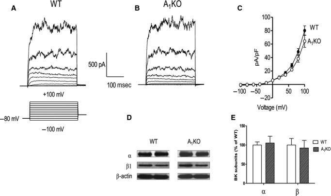
Whole-cell K+ current and BK channel subunit expression is similar in smooth muscle from wild type (WT) and A1KO mice. Representative traces of whole-cell K+ current in aortic smooth muscle cells from WT (A) and A1KO mice (B). The voltage template used to elicit the currents in this and subsequent figures is shown below the trace in A; cells were held at −80 mV and stepped from –100 to +100 mV in 20 mV increments. (C) Group data representing whole-cell K+ current in aortic smooth muscle cells from WT (n = 13) and A1KO (n = 20) mice. (D) Representative Western blots from mouse aortae for BK channel subunit expression relative to β -actin (α = 100 kDa; β 1 = 25 kDa; β –actin = 42 kDa). (E) Group data for BK α and β 1 subunit expression in the aortae of WT (n = 6) and A1KO (n = 6) mice. There were no differences between WT and A1KO mice in whole-cell K+ current or BK protein expression.