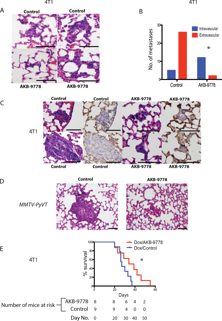
Figure 4.
Effect of vascular endothelial protein tyrosine phosphatase (VE-PTP) inhibition on extravasation of disseminated tumor cells into distant organ parenchyma. A–C) 4T1 mammary carcinoma cells were injected intravenously, and therapy with control or AKB-9778 commenced 12 hours later. Lungs were examined for micrometastases after 8 days. A) Representative hematoxylin and eosin–stained sections of micrometastases. Control lungs (upper panels) show micrometastatic cells that have breached vascular boundaries and entered the alveolar airspace. AKB-9778–treated lungs (lower panels) show cells tracking within vessels yet to extravasate (scale bars = 100 µm). B) Number of intra- vs extravascular micrometastases (*P < .01 by two-sided Fisher exact test; n = 6 mice per group; data show total number of metastases combined for all mice in each group). C) Representative images of micrometastases. Images show hematoxylin and eosin–stained sections of micrometastases and serial sections (separated by 5 microns) showing the same metastasis stained for collagen IV (brown). Control mice show extravasation of tumor cells through the vascular basement membrane, but AKB-9778–treated mice show tumor cells retained within vessels (scale bars = 100 µm). D) Representative images of experimental MMTV-PyVT tumor cell micrometastases treated with control (left panel) or AKB-9778 (right panel) (scale bars = 100 µm). E) Mouse survival after adjuvant therapy with doxorubicin ± AKB-9778. (*P = .05 using log-rank [Mantel–Cox] test; n = 8–9 mice per group) using a model of 4T1 spontaneous metastasis.

