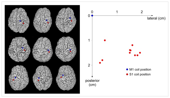
Figure 2. S1 coil target location in in nine representative subjects of experiment S1.
Left panel. The S1 target was identified using a custom MRI-guided neuronavigation system. The position of the coil was adjusted to target the post-central gyrus at a location mirroring the M1 hotspot relative to the central sulcus, i.e. the location expected to correspond to the representation of the hand within S1. The M1 (blue) and S1 (red) targets are shown on the cortical surface reconstructed from the individual MRI data of nine representative subjects. Right panel. Using our MRI-guided approach to target S1, we found that the actual location of the coil on the scalp surface was both more posterior and more lateral relative to the M1 coil position (x-axis: medial-lateral distance relative to the M1 coil position; y-axis: anterior–posterior distance relative to the M1 coil position).

