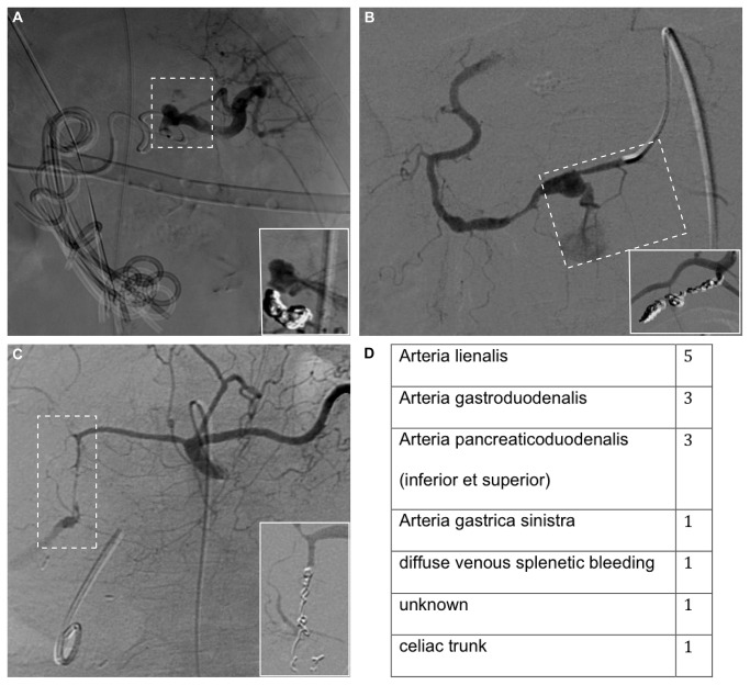
Figure 1. Illustration of bleeding vessels before and after transcatheter arterial coil embolization.
Digital subtraction angiography showing bleeding of arteria lienalis (A), arteria pancreaticoduodenalis inferior (B), and arteria gastroduodenalis. Inlets demonstrate transarterial coil embolization. The localizations of intraabdominal bleedings are shown in the table (D).

