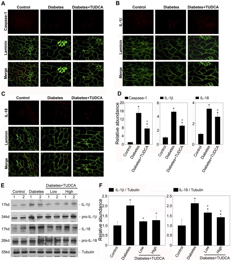
Figure 9. TUDCA attenuates the inflammasome activation in STZ induced diabetic nephropathy.
A, B and C: Kidney cryosections were stained by an indirect immunofluorescence technique. Representative micrographs from different groups as indicated showed the immunostaining of inflammasome markers in diabetic nephropathy treated without (middle column) or with 500 mg/kg/day TUDCA (right column). D: The histogram demonstrated the relative abundance of the semiquantitative histomorphometric analysis of the immunostaining of caspase-1, IL-1β, and IL-18. E: Western blot analysis showed TUDCA suppressed the maturation of IL-1β and IL-18 protein in STZ induced diabetic nephropathy. The kidney lysate (made from the pool of kidneys from six animals/group) were separated on a SDS-polyacrylamide gel and immunoblotted with a specific monoclonal antibody against IL-1β, IL-18 and α-tubulin, respectively. Samples from two individual animals were used at each time point. F: Quantitative determination of IL-1β and IL-18 protein abundance after normalization with α-tubulin. Data are presented as means ± SEM of three experiments. n = 6, *P<0.05 vs. normal control. #P<0.05 vs. diabetic group.

